


Arthropathy is a chronic pathology affecting the connective tissue structure of the musculoskeletal system.The disease is characterized by a progressive course with gradual destruction of cartilage tissue.Most patients do not discover joint disease until they are 65 years old, as the body's natural aging plays a role in its development.
The development of degenerative dystrophic pathologies is caused by previous injuries, endocrine and inflammatory diseases, excessive physical activity or conversely a sedentary lifestyle.The main symptoms of arthrosis are joint pain, swelling and limited movement.
To diagnose pathology, instrumental studies are required - radiography, arthroscopy, MRI, CT.Arthropathy of first and second severity can be treated conservatively with a range of medications, physical therapy and massage procedures, and exercise therapy.If irreversible destructive changes occur in the joint, surgical intervention is required - arthrodesis, endoprosthesis.
Pathogenesis
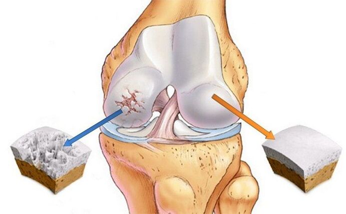
With arthropathy, significant changes occur in the internal connective tissue structure.Deformable erosions form on the cartilage tissue, leading to the destruction of collagen fibers and proteoglycans composed of proteins (5–10%) and glycosaminoglycans (90–95%).As a result, the collagen network loses stability and metalloproteinases begin to be released, destroying all types of extracellular matrix proteins.Accelerates destruction by increasing biosynthesis of collagenase and stromelysin.Normally, the normal quantitative values of enzymes are controlled by cytokines (small peptide messaging molecules).But as arthrosis progresses, the concentration of these proteins decreases, triggering the release of large amounts of enzymes that damage cartilage tissue.
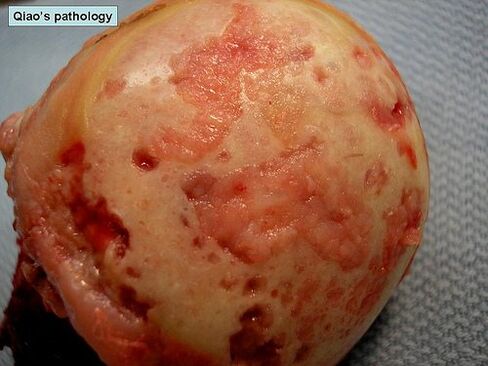
The structurally altered proteoglycans begin to absorb water molecules that they cannot retain.Therefore, excess fluid enters the collagen fibers.They "swell" and lose strength and elasticity.The qualitative and quantitative composition of synovial fluid also undergoes negative changes.As arthropathy develops, the concentration of hyaluronic acid in it decreases.The hyaline cartilage no longer receives enough nutrients and oxygen to regenerate.Foci of softening form in the cartilage tissue, followed by cracks and specific necrotic growth.As bones shift relative to each other, the bones are exposed and begin to suffer microtrauma.
Causes and Predisposing Factors
The cause of primary (idiopathic) arthropathy has not been determined.It occurs in the absence of any predisposing factors, leading to theories regarding a genetic predisposition to premature cartilage destruction.Secondary arthropathy is caused by other joint pathology or previous injury.What may cause degenerative dystrophic diseases:
- Damage to joints or nearby connective tissue structures - fractures, dislocations, meniscus injuries, partial rupture of muscles, ligaments, tendons or complete separation from the bone base;
- congenital joint dysplasia;
- Endocrine gland dysfunction and metabolic process disorders;
- Rheumatism or rheumatic fever;
- Rheumatoid arthritis, reactive arthritis, metabolic arthritis, psoriasis or gouty arthritis, polyarthritis;
- Septic arthritis caused by Streptococcus, Epidermidis, or Staphylococcus aureus;
- Tuberculosis, brucellosis, chlamydia, gonorrhea, syphilis of any site;
- Degenerative diseases such as osteochondritis dissecans.
Excessive joint movement due to the production of special collagen can easily lead to the occurrence of arthropathy.The disease affects 10% of the planet's inhabitants but is not considered a pathology.But excessive activity is accompanied by weakness of the tendon-ligament mechanisms, which can lead to frequent injuries, especially of the ankle joint (ligament sprains and ruptures, dislocations).
Osteoarthritis is sometimes caused by disorders of the blood-forming system, such as hemophilia.Hemarthrosis or bleeding into the joint cavity can cause deterioration of cartilage nutrition and its destruction.
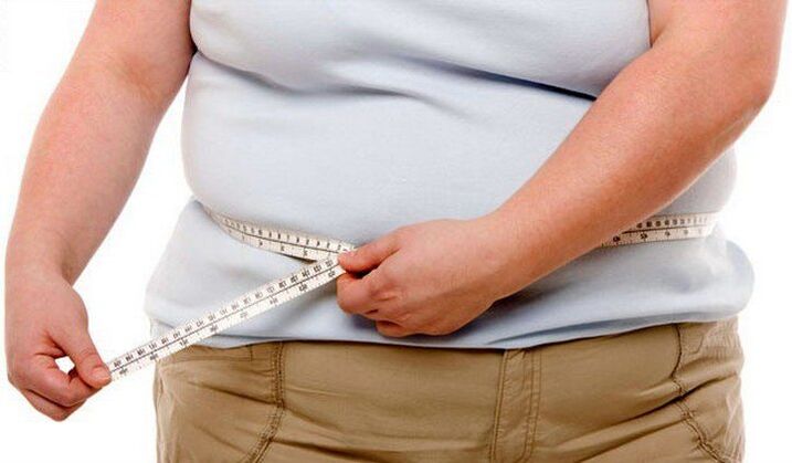
Predisposing factors include old age, frequent loading of joints beyond their strength limits, excess body weight, surgical intervention, and hypothermia.
Risk groups include menopausal women, people living in adverse environmental conditions or exposed to toxic compounds.A lack of vitamins and trace elements in the diet creates prerequisites for the gradual destruction of hyaline cartilage.
clinical picture
The danger of arthropathy is that there are no symptoms in the first stages of its development.The pathology manifests clinically gradually, the first signs appearing against the background of significant destruction of cartilage tissue.Initially, a person will feel mild pain, but it is not clearly localized.It occurs after physical activity - weightlifting, sports training.Sometimes the first clinical sign is a crunching or clicking sound when bending or extending the joint.A person begins to notice that some movements are difficult.However, in the initial stages of arthrosis, stiffness occurs in the morning, which quickly disappears.
As the condition progresses, pain may also be felt at night, causing not only sleep disturbance but also chronic fatigue.The severity of stage II pain syndrome increases with changes in weather, exacerbation of chronic disease, and acute respiratory viral infections.The range of activities is significantly reduced.Stiffness is caused by thinning of the cartilage and the person's conscious restriction of movement to avoid pain.This results in increased loads on the opposing joints, causing further damage.Arthropathy also has other specific symptoms:
- Pain causes skeletal muscle spasm and muscle contracture (limited passive movement of joints);
- The creaking, clicking, and snapping sounds of the joints as they move become constant and occur almost every time the bones move relative to each other;
- Frequent painful muscle spasms;
- Deformation of joints, resulting in disordered posture and gait;
- In the third stage of arthrosis, the deformation is very obvious, causing the joint to bend, and the range of motion of the joint is significantly reduced or completely eliminated;
- Patients with three-degree joints in the knees, ankles, and hips need to use canes or crutches when moving.
Without treatment, the disease progresses, and over the course of the disease remissions are replaced by relapses, with exacerbations increasing in frequency.The stiffness of movement in the morning now does not go away for a long time but is permanent.
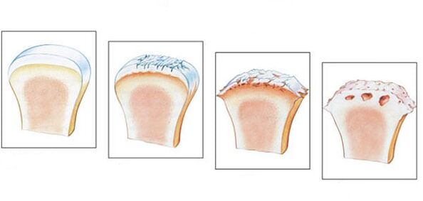
When examining a patient with grade 1 arthropathy, doctors note only mild swelling of the joint but complete preservation of range of motion.In grade 2 pathology, palpation reveals pain and mild deformity.In the joint space area, the formation of bone thickening was observed.
Arthropathy is characterized by the development of synovitis - an inflammatory process of the synovium of the hip, knee, ankle and shoulder joints.Their main symptoms are the formation of a circular seal in the joint area and the feeling of movement (fluctuation) of fluid when pressure is applied.Acute synovitis may be accompanied by an increase in body temperature to 37-38 °C, headache, and digestive disturbances.
diagnosis
Diagnosis is made based on instrumental findings, clinical presentation features, medical history, and patient complaints.General blood and urine tests do not provide much information - if the joint is not caused by metabolic pathology, all values remain within the normal range.As synovitis develops, the erythrocyte sedimentation rate increases (30 mm/h), and the levels of white blood cells and fibrinogen in the blood increase.This indicates an acute or chronic inflammatory process occurring in the body.Changes in biochemical and immunological parameters occur in secondary arthropathy.
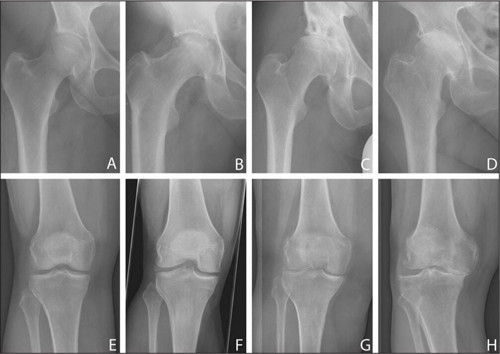
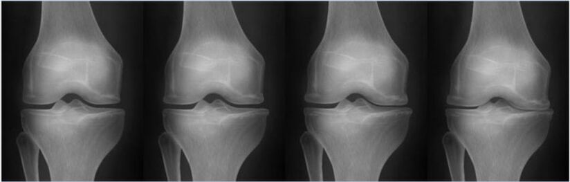
The most informative method for diagnosing degenerative dystrophic pathology is radiography in frontal and lateral projections.
| Staging of arthropathy according to the Kellgren-Lawrence classification (1957) | X-ray pathological signs |
|---|---|
| initial | No sign of radioactivity |
| First | The joint space is not obvious and narrows unevenly.Slight flattening of bone plate edges and initial osteophyte formation or disappearance |
| second | The joint space is significantly narrowed, 2-3 times higher than normal, a large number of osteophytes are formed, and the subchondral bone is sclerotic.Cystic spaces appear in the epiphysis |
| third | There is obvious subchondral bone sclerosis and large marginal osteophytes, and the joint space is significantly narrowed. |
| fourth | Thick osteophytes are formed, the joint space is almost completely fused, and the epiphysis forming the joint is deformed and compacted. |
A CT scan is done if the doctor has doubts about the diagnosis after studying the X-ray images.To evaluate the condition of the connective tissue structures near the joint, an MRI is performed.When using contrast media, it is possible to dynamically assess the blood supply of the tissue and determine the stage of the inflammatory process in the development of synovitis.
Basic treatment methods
Arthropathy remains an incurable disease because there are no drugs that can regenerate cartilage tissue.The main goals of treatment are to prevent progression of pathology and maintain joint range of motion.Treatment is long-term, complex, and requires the use of topical and systemic medications.Patients should avoid excessive stress on their joints and, if necessary, use orthopedic devices (orthotics, elastic bandages) to limit range of motion.Overweight patients need to adjust their diet, gradually lose weight, and follow dieting principles.
After achieving stable remission, the patient received daily physical therapy exercises.The first training session is conducted under the supervision of a physiotherapist, and then the patient performs a set of exercises at home.Exercise therapy can be supplemented with swimming, yoga and cycling.
To reduce the severity of pain, drugs from various clinical and pharmacological groups are prescribed:
- Non-steroidal anti-inflammatory drugs in the form of ointments, tablets, parenteral solutions containing active ingredients;
- A combination of anesthetic solution and corticosteroids is injected into the joint;
- Muscle relaxants relieve muscle spasms and restrictive contractures.
Treatment options include B vitamins, sedatives, and, if necessary, sedatives and antidepressants.Chondroprotectants are required for long-term use.This is the only group of drugs that can partially restore cartilage tissue.
To increase their clinical activity, physical therapy procedures are performed - laser therapy, magnetic fields, ultrahigh frequency therapy.
Any pain in the joints should be a sign to consult a doctor immediately.Treatment during the initial stages of arthrosis will halt the destruction of cartilage and avoid loss of function and disability.