


What is spinal osteochondrosis in simple terms?
Spinal osteochondrosis is a chronic disease based on degenerative dystrophic changes in the intervertebral discs, subsequently involving adjacent vertebrae, intervertebral joints, and spinal ligaments in the process.
The word "osteochondrosis" has two Greek roots: οστό - bone and χόνδρος - cartilage.
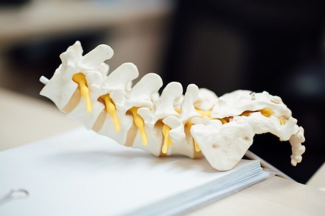
Vertebrae are structures made of cancellous bone.They are connected to each other by discs of cartilage.There are ligaments along the front and back surfaces of the vertebrae.Disks of cartilage prevent the vertebrae from coming together and the ligaments from moving.The spine is elastic due to the coordinated work of the discs and ligaments, which allows it to perform important functions:
- Ensure vertical position balance,
- Reduces shock and vibration when walking and jumping,
- Protects the skull and the brain located within it from shock caused by excessive impact.
In osteochondrosis, the disc herniates beyond the vertebral body.Depending on the direction in which the protrusion occurs and its size, pain, numbness, muscle confusion, and other symptoms may occur.
ICD-10 code:
- M42 Spinal Osteochondrosis
- M42.0 Young osteochondrosis of the spine
- M42.1 Adult spinal osteochondrosis
- M42.9 Spinal osteochondrosis, unspecified
- M43.1 Spondylolisthesis
- M47 Spondylosis
- M47.0 Anterior spinal artery or vertebral artery compression syndrome
- M47.1 Other spondylosis associated with myelopathy
- M47.2 Other spondylosis with radiculopathy
- M48.0 spinal stenosis
- M50.0 Cervical disc injury with myelopathy
- M50.1 Cervical disc injury with radiculopathy
- M50.2 Another type of cervical disc displacement
- M50.3 Other cervical disc degeneration
- M51.0 Lumbar disc disease and other myelopathy
- M51.1 Lumbar disc disease and other radiculopathy
- M51.2 Other specified disc displacements
- M51.3 Other specified disc degeneration
- M53 Other back disorders not classified
Types of osteochondrosis
There are several variants of this disease depending on which part of the spine the changes occur:
- cervical spine,
- Chest,
- lumbar spine,
- sacrum,
- Mixed variant (cervicothoracic, lumbosacral).
Depending on the duration of symptoms, the disease can be:
- Acute (up to 3 weeks),
- Subacute (3-12 weeks),
- Chronic (more than 12 weeks).
According to the main neurological manifestations:
- Have myelopathy (spinal cord injury),
- Have radiculopathy (compression and inflammation of nerve roots).
Causes of osteochondrosis
To date, there are no firm data on the causes of osteochondrosis.
The role of genetic predisposition, mechanical injury, and inflammation in the development of premature disc wear is recognized.
The intervertebral discs do not have their own blood or lymphatic vessels.The blood vessels of the vertebrae play a role in nourishing and removing harmful substances.As we age and/or are exposed to harmful influences, blood and lymph flow decreases, the discs receive less oxygen and nutrients, and harmful substances can accumulate in them.All of this leads to gradual wear and tear.The extent and rate of brake disc wear increases when exposed to hazardous factors.
Risk factors:
- Congenital anomalies of the vertebrae and spinal canal;
- flatfoot;
- Occupational hazards (vibration, lifting heavy objects, prolonged exposure to forced uncomfortable positions, exposure to toxic substances);
- sedentary lifestyle;
- obesity;
- a diet that is unbalanced in protein, fat, vitamins, and minerals;
- Inadequate consumption of clean water;
- smoking;
- environmental pollution.
Symptoms of spinal osteochondrosis
Listed by frequency of occurrence:
- pain;
- reduced range of motion;
- numbness, loss of sensitivity;
- Decreased muscle strength;
- Innervation of organ dysfunction associated with problem areas in the spine.
Clinically significant manifestations of spinal osteochondrosis were observed in 51 per 1000 persons.
The location of pain and other symptoms depends on the problem area of the spine.
Cervical osteochondrosis:
- Pain in arms, shoulders, and neck that is aggravated by turning and tilting the head;
- Headache;
- Decreased arm muscle strength;
- Noise in the head, dizziness, "floaters", spots in front of the eyes with burning sensation, throbbing headache (vertebral artery syndrome).
The health of the brain depends on the condition of the cervical spine because the arteries to the brain pass through tubes formed by the protrusions of the vertebrae.If the lumens become narrowed due to osteochondrosis, blood flow through the arteries is disrupted and the brain becomes starved of oxygen and nutrients.
Osteochondrosis of the chest:
- Pain in the chest, under the shoulder blades, and in the heart area, worsened when turning the body, coughing, or sneezing;
- Gallbladder, stomach, and esophagus dysfunction.
Osteochondrosis of the lumbar spine and/or sacrum:
- Pain in the lower back, back of the thighs, and sides;
- numbness in toes;
- Increased frequency of urination (10-12 times per day, possibly more), involuntary leakage of urine during physical activity;
- Sexual disorders.
Half of people with osteochondrosis show signs of ongoing emotional stress due to frequent pain.
Development stages and course of osteochondrosis
The initial symptoms of osteochondrosis include dull pain in the back or waist after standing, walking, or running for a long time; neck pain that is aggravated by turning and tilting the head.
As disc disease progresses, the disc may bulge (herniate), compressing the nerve root (radiculopathy).This can lead to severe pain in the arms or legs, muscle weakness, skin sensitivity, blood vessel tone, and dysfunction of the organs receiving innervation from the problem area of the spine.In the most severe cases, compression of the spinal cord may occur, resulting in paresis or paralysis.
Osteochondrosis is a chronic disease.After adequate treatment, remission occurs, which is when symptoms decrease or disappear completely.If a new herniation develops in the disc, the condition may worsen and pain and other symptoms may return.
diagnosis
Examination by a neurologist.
Basic instrumental research methods:
- Magnetic Resonance Imaging (MRI),
- Computed tomography (CT).
Additional:
- Myelogram (an in-depth X-ray examination of the spine),
- Electromyography (EMG),
- Electromyography (ENMG),
- Bone densitometry (to detect osteopenia/osteoporosis).
Basic laboratory methods:
- General blood tests,
- General urinalysis,
- Blood biochemical tests (blood sugar, creatinine, urea, electrolytes, bilirubin, liver enzymes and pancreatic enzymes; glycosylated hemoglobin, C-reactive protein),
- Coagulation diagram.
Additional:The concentration of calcium and phosphate in the blood.
Treatment of osteochondrosis
Conservative treatment
This test is performed if the patient does not have acute progressive neurological symptoms.
Target:
- Reduce or relieve pain,
- Correct muscle tone,
- Reduce inflammation and swelling,
- Prevents the progression of dystrophic changes in spinal structures,
- Correct damaged functions of internal organs,
- Increase patients’ daily activities,
- Teach patients to cope with pain.
Conservative treatment of osteochondrosis includes:
- Follow a reasonable exercise regime,
- using drugs,
- physical therapy,
- massage,
- exercise therapy (after pain relief and stabilization),
- acupuncture,
- Manual therapy.
drug treatment
Lists the main groups of drugs that relieve or reduce pain and stabilize the condition of patients with osteochondrosis.Only a physician can consider the characteristics of a specific patient's clinical situation and select an appropriate treatment plan.
NSAIDs(NSAIDs):
- For oral administration,
- for intramuscular injection,
- For intravenous administration,
- for insertion into the rectum (rectal suppositories),
- Topical (ointment, gel).
muscle relaxants(medication to reduce muscle spasms).
For severe tightness and painful muscle spasms.
diuretics(Reduce local swelling).
Drugs that improve the condition of cartilage tissue(chondroprotectant):
- Chondroitin sulfate sodium,
- A combination of chondroitin sulfate sodium and glucosamine.
B vitamins:
- Thiamine (B1),
- Pyridoxine (B6),
- Cyanocobalamin (B12),
- Combination B1+B6+B12.
If the pain is severe in the acute stage, you can rest in bed for 1-2 days, which can help relax the muscles and reduce the pressure in the cartilage disk.A waist stabilizing corset or Shants collar is recommended.
As the pain intensity decreases, treatment is supplemented with special therapeutic exercises aimed at stretching the spine and relaxing the muscles, and gradually includes exercises to form a corset of the muscles.Therapeutic manual massage is indicated.
With adequate treatment, the pain will gradually lessen and may even disappear completely.Neurological symptoms also subsided.Improvement is due to a reduction in the size of the herniated disc and associated inflammatory changes in the surrounding tissue.
surgical treatment
Urgent neurosurgical intervention is indicated for pelvic disease associated with numbness of the anogenital area and ascending paralysis of the feet (cauda equina syndrome).
Surgery may also be needed if conservative treatments fail within 3-6 months.
Prevent back pain
Avoid excessive physical activity (lifting heavy objects, carrying heavy bags in one hand, etc.).
Avoid prolonged static loading (sitting, maintaining uncomfortable positions).
If your job involves such stress, it is recommended that you take a 10-minute break every 45 minutes and walk during this time.
Avoid hypothermia.
Maintain an appropriate level of physical activity through regular exercise, swimming and/or walking.
Sleep on a medium-firm mattress.
Nutrition for osteochondrosis
A balanced diet and proper fluid intake ensure normal blood supply and nutrients to the vertebrae as well as the cartilaginous discs.As a result, metabolism and energy are normalized and harmful products do not accumulate.
Basic principles:
daily calories, calculated separately, taking into account height, age, and gender.
For patients who are overweight or obese, caloric intake should be restricted.
drinking habits– Drink at least 1 liter of purified water, mineral water and herbal tea every day, preferably 30 ml per kilogram of body weight.
Daily use:
- Whole grain products (buckwheat, millet, oats);
- Adequate amounts of protein (taking into account age and kidney function): animals - lean beef, chicken, turkey, rabbit, eggs (4-5 per week); vegetables - beans, lentils, peas;
- Healthy fats containing monounsaturated and polyunsaturated fatty acids (fish, seafood, unrefined vegetable oils, unrefined and unsalted nuts, seeds);
- Vegetables (fresh and cooked), lettuce, herbs and leafy greens;
- Berries - blueberries, blackberries, raspberries, cherries.
Eliminate from the diet:
- white bread and bakery products made from high-quality flour;
- Sugar, industrial confectionery - candies, cakes, biscuits, gingerbread, waffles;
- Industrial beverages with added sugar - carbonated water, packaged juices;
- Processed meat products – sausages, salami, canned foods.