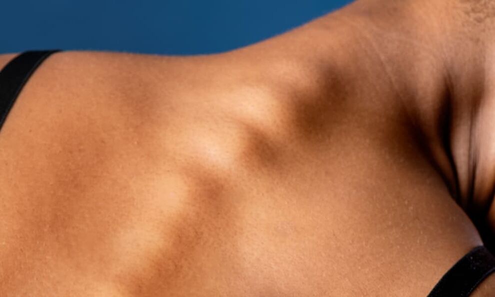
Causes of osteochondrosis
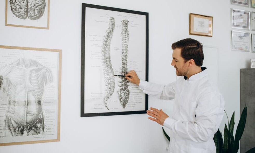
- genetics,
- Physical development defects,
- metabolic diseases,
- unhealthy diet
- Lack of vitamins and minerals,
- long-term use of medications,
- overweight,
- Increase the load on the spine,
- A sedentary lifestyle, such as when working in an office,
- spinal injury,
- Past infectious diseases and stress.
Signs and symptoms of osteochondrosis
- Pain in various parts of the body, such as in the back, neck, or other areas;
- Difficulty and restriction of movement when turning or bending;
- persistent tension and muscle spasms;
- Migraines and dizziness;
- Pain in the heart area;
- Muscular hypotension, decreased muscle tone and strength;
- numbness in limbs;
- pain in arms and legs;
- Spots appear before eyes;
- cooling of extremities;
- Photograph the painful feeling.
- loss of consciousness;
- Decreased sensitivity of limbs;
- Poor blood circulation in blood vessels;
- nerve damage or inflammation;
- Arteries become narrowed and blocked.
risk factors
- Nervous and emotional exhaustion;
- physical overexertion;
- working on a vibrating platform;
- genetic susceptibility;
- Lack of vitamins in the body;
- Multiple pregnancy.
Classification and stages of development of osteochondrosis
Classification of osteochondrosis
- Lombardinha– This is lumbar (lumbosacral) back pain.
- sciaticaPresents as pain in the back that spreads to the legs.
- low back pain——This is lumbago, acute, severe pain in the waist.
- chest pain- This is pain in the chest.
Stages of development of osteochondrosis
- I.In the first stage, the disc core loses moisture and becomes less elastic, leading to loss of height and tissue breakdown. During this stage, the pain is usually barely noticeable, but discomfort may occur during physical activity or abnormal postures.
- two.In the second stage of osteochondrosis development, the disc tissue begins to flatten and bulge, causing the spaces between the vertebrae to narrow and pinching the spinal nerve roots. The fibrous membrane is destroyed, resulting in poor fluid retention in the disc core. You can hear characteristic clicking and crunching sounds in your spine as you move. During this stage, some pain may occur and worsen with active movement.
- three.Stage three is characterized by wear and thinning of the cartilage lining between the discs. At this stage, symptoms of osteochondrosis manifest themselves in the form of acute pain. For quick pain relief, nerve pain painkillers are taken.
- Four.In the final, fourth stage, the discs are so damaged that the joints become immobile and the spaces between the vertebrae become overgrown with bone tissue. Severe dystrophic processes can cause acute pain as growth damages adjacent tissue and compresses nerves. The mobility of the vertebral joints may be completely lost.
complication
- herniated disc, which occurs when the nucleus pulposus of an intervertebral disc herniates beyond the annulus fibrosus. This can lead to spinal pain and dysfunction.
- intervertebral hernia- This is a more serious complication when the disc annulus ruptures and the nucleus pulposus extends beyond it. This can cause severe pain, decreased sensation, and paralysis.
- Radiculitis- This is nerve root compression with severe pain symptoms. Radiculitis can cause loss of sensation, numbness, and weakness in the lower extremities.
- KyphosisIt is a spinal deformity that manifests as a bulge in the chest. This can lead to breathing problems, pain, and poor posture.
- spinal cord stroke– This is the most serious complication of osteochondrosis and can lead to loss of sensitivity, impaired motor function, and even paralysis.
- Lower limb muscle atrophy– This is a loss of muscle mass, accompanied by rapid fatigue and weakness in the legs.
- Leg paralysis– This is a complete loss of voluntary movement of the lower limbs, a serious complication of osteochondrosis.
How to diagnose osteochondrosis
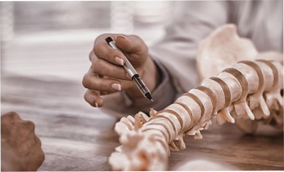
- Physical examination.The doctor examines the patient and assesses his general condition, posture, and movements. Doctors may also perform neurological tests to determine whether sensory and motor problems are present.
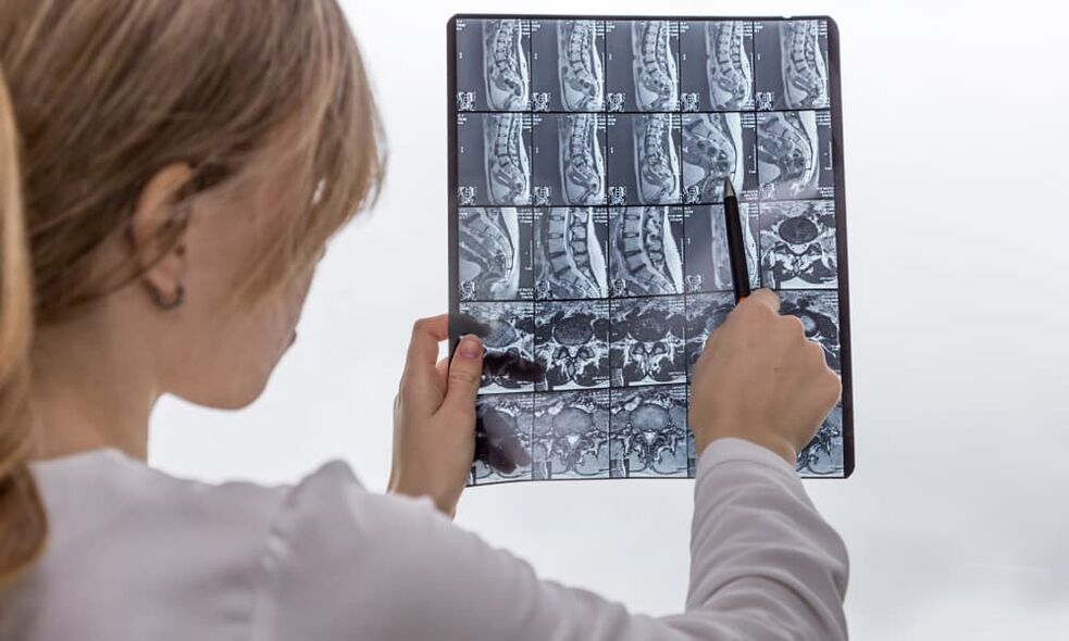
- Hardware check.For more accurate diagnosis, various hardware examination methods are used, including radiography, computed tomography (CT), and magnetic resonance imaging (MRI).
- blood test.A complete blood count can help identify early symptoms of osteochondrosis, such as an increased erythrocyte sedimentation rate and low calcium levels. To confirm the diagnosis, biochemical tests can be performed to evaluate coagulation parameters, enzyme activity, levels of zinc, cobalt, iron and other components.
- Radiography.During an X-ray, each spine is examined and pictures are taken in direct, lateral, and two oblique projections. If necessary, functional radiographs may be performed to allow you to evaluate the condition of your spine in different locations.
- Computed tomography (CT). A CT is performed after an X-ray and allows you to more accurately determine the condition of the disc. To do this, a photo of one or two segments of the spine is taken.
- Magnetic resonance imaging (MRI).MRI may be used as a supplement to CT or in situations where more detailed study of blood vessels, nerve processes, and disc conditions is required.
When to see a doctor
Treatment of osteochondrosis
exercise therapy
Using manual therapy to treat osteochondrosis
massage
- Puts extra stress on muscles and increases their tone;
- Eliminate lactic acid accumulation and relieve muscle spasms;
- Improves blood circulation to the affected area and adjacent tissues;
- relief the pain.
physiotherapy
- Magnet therapyIt is the effect of a constant frequency magnetic field that stimulates cellular responses.
- Osteochondrosis electrophoresis– This is the effect of an electric field on the tissue, which accelerates blood circulation and activates regenerative processes.
- Laser TreatmentIt is a method of stimulating the biological processes of nerve fibers and also has anti-inflammatory, wound healing and analgesic effects.
- shockwave therapyIt is a method of using sound waves to affect diseased parts of the body, which can improve microcirculation and metabolic processes and relieve swelling and pain.
Kinesio tape
acupuncture
Leech therapy
medical treatement
reflexology
Prevention and prognosis of osteochondrosis

- genetic predisposition to spinal disease;
- Chronic gastrointestinal problems may lead to malabsorption of nutrients;
- Diseases associated with metabolic disorders;
- Having serious infections in childhood, such as rickets;
- spinal injuries;
- overweight.
osteochondrosis diet
Exercises for osteochondrosis
- perform head tilt;
- Turn your head from side to side;
- Use your chin to draw numbers from 0 to 9 in the air;
- Move your chin back and forth in a horizontal plane.


